
Etiological and Histopathological Spectrum of Granulomatous Skin Diseases: A Retrospective Descriptive Study from a Tertiary Care Hospital in Pakistan
DOI:
https://doi.org/10.66344/jpad.v36i2.3198Keywords:
etiological spectrum, histopathology, granulomatous diseasesAbstract
Background Granulomatous skin diseases include diverse groups of disorders, and they pose a diagnostic difficulty because of their overlapping clinical features. Although they have a wide etiological spectrum, from infectious diseases like tuberculosis to noninfectious diseases like sarcoidosis. Due to high prevalence of these infections in Pakistan, accurate histopathological evaluation is needed to guide proper treatment.
Objective To find out the etiological spectrum, prevalence, and histopathological patterns of granulomatous diseases and to evaluate their regional distribution and clinical significance.
Methods This retrospective descriptive cross-sectional study was carried out between January 2021 and December 2023 at Khyber Teaching Hospital, Peshawar. Among them, 60 cases (16.7%) with confirmed histopathological granulomatous inflammation were included. Immunohistochemistry, and special stains were used to confirm the specific aetiology where needed. Data were analyzed using SPSS 22.0.
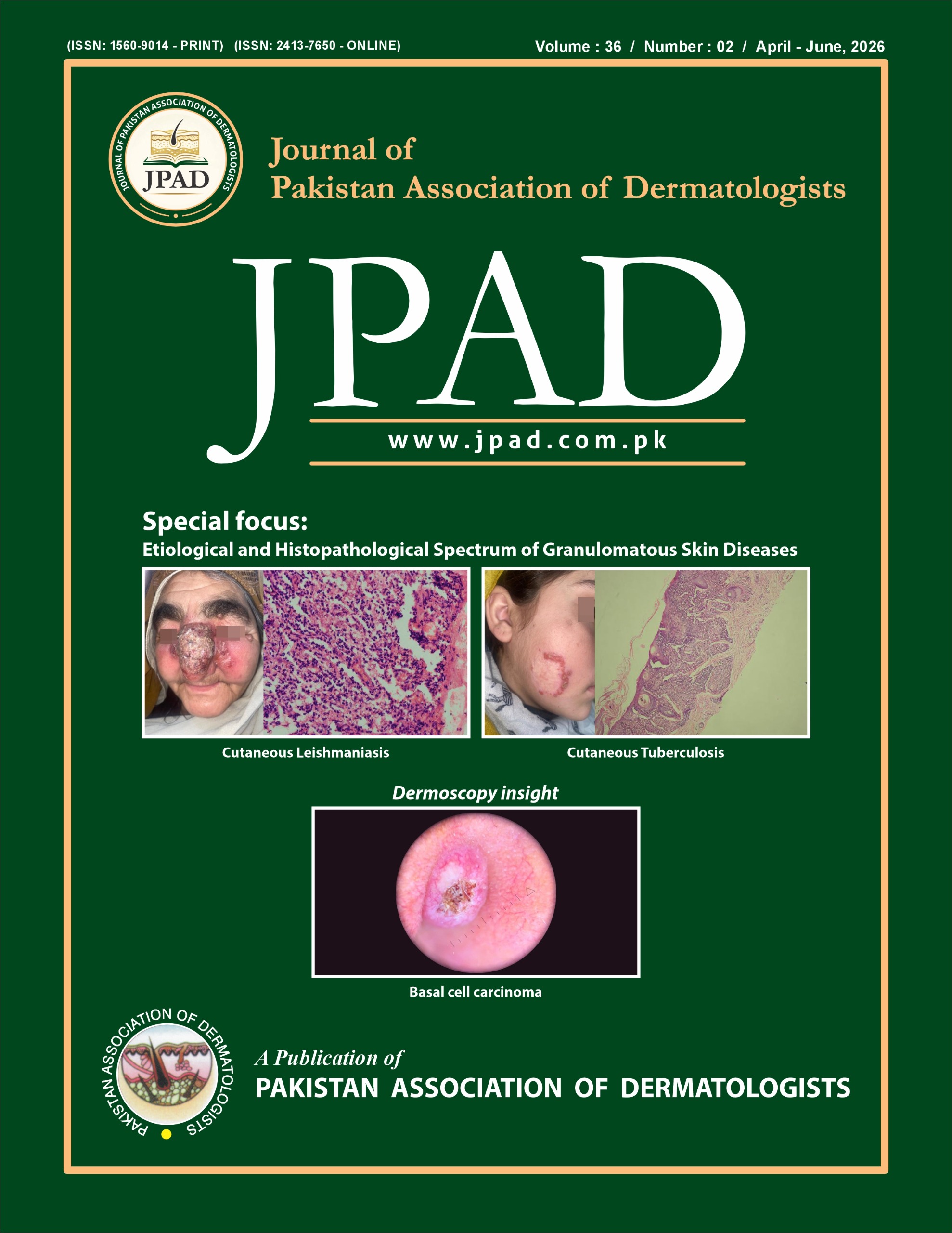
Results Infectious etiologies were most common, led by cutaneous tuberculosis (38.4%, n=23) followed by cutaneous leishmaniasis (33.4%; n=20), leprosy (10%; n=6) and syphilis (5%; n=3). Among the noninfectious cases, sarcoidosis was on top (8.3%; n=5) followed by foreign body granuloma (5%; n=3). Lupus vulgaris was the most predominant subtype of cutaneous tuberculosis (65.2%; n=15) followed by scrofuloderma (26%; n=6) and Tuberculosis verrucosa cutis (8.69%; n=2). Lepromatous Leprosy was the most frequently observed form of leprosy.
Conclusion Granulomatous diseases represent a significant health burden in Pakistan, particularly cutaneous tuberculosis and cutaneous leishmaniasis which account for over 70% of cases. Early skin biopsies with a clinicopathological correlation and aided by special stains and immunohistochemistry where needed is essential for accurate diagnosis, proper treatment, and improved patient outcome.
References
1. Tronnier M, Mitteldorf C: Histologic features of granulomatous skin diseases. Part 1: non-infectious granulomatous disorders. J Dtsch Dermatol Ges. 2015;13(3):211-6.
2. Kumar L, Agarwal P, Mishra T, Chahar Y, Kamal R, Tyagi S, et al: Study of histomorphological spectrum of granulomatous lesions of skin. Tuberculosis. 2021;57:44-56.
3. Terziroli Beretta-Piccoli B, Mainetti C, Peeters MA, Laffitte E: Cutaneous granulomatosis: a comprehensive review. Clin Rev Allergy Immunol. 2018;54(1):131-46.
4. George VP, Srinivasan S: Histopathological spectrum of granulomatous skin lesions: a review. SBV J Basic Clin Appl Health Sci. 2019;2(3):95-104.
5. Asai J: What is new in the histogenesis of granulomatous skin diseases? J Dermatol. 2017;44(3):297-303.
6. Wick MR: Granulomatous and histiocytic dermatitides. Semin Diagn Pathol. 2017;34(3):301-11.
7. Chatterjee D, Bhattacharjee R, Saikia UN: Non-infectious granulomatous dermatoses: a pathologist’s perspective. Indian Dermatol Online J. 2021;12(4):515-28.
8. Lehman JS, Sokumbi O, Peters MS, Bridges AG, Comfere NI, Gibson LE, et al: Histopathologic features of noninfectious granulomatous disorders involving the skin. Hum Pathol. 2020;103:127-45.
9. Chopra A, Mitra D, Sharma L, Agarwal R: Granuloma annulare skin lesions in a case of sarcoidosis. Indian Dermatol Online J. 2018;9(2):117-9.
10. Zafar MN, Sadiq S, Memon MA: Morphological study of different granulomatous lesions of the skin. J Pak Assoc Dermatol. 2008;18(1):21-8.
11. Khanna V, Goel N: Histopathological approach to granulomatous skin lesions. Indian J Dermatol Venereol Leprol. 2010;76(1):48-56.
12. Chakrabarti S, Pal S, Biswas BK, Bose K, Pal S, Pathak S: Clinico-pathological study of cutaneous granulomatous lesions: a 5-year experience in a tertiary care hospital in India. Iran J Pathol. 2016;11(1):54-9.
13. Pawale J, Belagatti SL, Naidu V, Kulkarni MH: Histopathological study of cutaneous granuloma. Indian J Public Health Res Dev. 2011;2(2):74-9.
14. Dhar S, Dhar S: Histopathological features of granulomatous skin diseases: an analysis of 22 skin biopsies. Indian J Dermatol. 2002;47(2):88-90
15. Gautam K, Pai RR, Bhat S: Granulomatous lesions of the skin. J Pathol Nepal. 2011;1(2):81-6.
16. Lin IT, Gin TH, Wen KJ: Spectrum of cutaneous granulomatous lesions: a 5-year experience in a tertiary care centre in Sarawak. Med J Malaysia. 2023;78(2):185-90.
17. de Brito AC, de Oliveira CM, Unger DA, Bittencourt MD: Cutaneous tuberculosis: epidemiological, clinical, diagnostic and therapeutic update. An Bras Dermatol. 2022;97(2):129-44.
18. Tobin EH, Warda K, Gropper C, Apple A, Vadakekut ES: Cutaneous tuberculosis. StatPearls [Internet]. Treasure Island (FL): StatPearls Publishing; 2025.
19. Singal A, Sonthalia S: Cutaneous tuberculosis in children: the Indian perspective. Indian J Dermatol Venereol Leprol. 2010;76:494-503.
20. Bhutto AM, Solangi A, Khaskhely NM, Arakaki H, Nonaka S: Clinical and epidemiological observations of cutaneous tuberculosis in Larkana, Pakistan. Int J Dermatol. 2002;41(3):159-65.
21. Ahmad S, Zafar N, Qurni MO, Orakzai SA, Khalil MA, Khan WA: Non-caseating granulomas in skin biopsies of Leishmania cases. J Islam Int Med Coll. 2023;18(1):15-21.
22. Balaram G: Histomorphological analysis of cutaneous granulomatous lesions. Dissertation (MD Pathology). Rajiv Gandhi University of Health Sciences, Bengaluru, India; 2011.
23. Chauhan P, Adya KA: Dermatoscopy of cutaneous granulomatous disorders. Indian Dermatol Online J. 2021;12(1):34-44.
24. Aubart FC, Ouayoun M, Brauner M, Attali P, Kambouchner M, Valeyre D, et al: Sinonasal involvement in sarcoidosis: a case-control study of 20 patients. Medicine (Baltimore). 2006;85(6):365-71.
25. Ishihara M, Ohno S: Genetic influences on sarcoidosis. Eye (Lond). 1997;11(2):155-61.
Downloads
Published
Issue
Section
License
Copyright (c) 2026 Journal of Pakistan Association of Dermatologists

This work is licensed under a Creative Commons Attribution 4.0 International License.
Submission declaration
Authors retain the copyright to their work and grant the 'Journal of Pakistan Association of Dermatologists (JPAD)' the right of first publication under a Creative Commons Attribution 4.0 International (CC BY 4.0) license. This license allows others to share, adapt, and reuse the work for any purpose, including commercial use, as long as appropriate credit is given to the original authors and the journal.
By submitting a manuscript, authors confirm that the work has not been published previously (except as an abstract, lecture, or academic thesis), is not under review elsewhere, and has been approved by all authors and relevant authorities. Once accepted, the article will be openly accessible under the CC BY 4.0 license, ensuring wide dissemination and reuse with proper attribution.


